Another Mycology Infodump:) - Tumblr Posts
Small staghorn - Calocera cornea
This post will discuss the small staghorn or the Calocera cornea. It is important to note that the description paragraph at the top of this post is a more generalised description, other paragraphs will go into more detail.

Description
Calocera cornea can be found on the deadwood of deciduous broadleaf hardwood trees, especially oaks, after heavy rains. On this deadwood it emerges in groups of smooth and cylindric fruiting bodies with rounded-off, or sometimes sharper tips.¹
While this fungus looks more like a club fungus than a jelly fungus, it is in fact a jelly fungus. This fungus namely has more gelatinous flesh instead of it being brittle. Additionally, microscopic examination shows this fungus to have Y-shaped basidia that charachterise members of the Dacrymycetales - a large class within the jelly fungi. ²
Fruitbody, spores and microscopic features.
The small staghorn can be found clustered in non-merging groups.² This fungus has a cylindric fruiting body with a rounded or sharpened apex (tip), which occasionally is shallowly forked. It grows about 2 - 18 mm high and 1 to 2 mm thick. The surface of the body is bald and greasy¹, firm but gelatinous.² Its coloration ranges from orange to orangish yellow, depending on weather circumstances and age. In drier weather the fruiting body will turn more orange and with age the apex will shrivel and get an orangish brown hue. This fungus has white mycelium.¹
Calocera cornea's smooth spores are ellipsoidal to sausage-shaped and about 7-10 x 2.5-4um in size. The spores are hyaline and inamyloid (clear and not changing colour with Melzer's reagent). When fully mature, they will often become 1-septate (developing a single cross wall), like seen in the picture on the left below. The basidia are Y-shaped or two-spored, visible in the picture on the right below.²

Ecology and biological role
This species is saprobic, growing scattered to clustered on the deadwood of decidious broadleaf hardwoods. They are mostly found on sticks up to five cm in diameter.¹
The species is saprobic, meaning that it feeds on dead or decaying organic matter. The small staghorn grows on deadwood, making its biological role to decompose wood.³
Distribution
The small staghorn is widely distributed in Europe (it is quite common in Britain and Ireland²), North America, Central America, South America, Asia, and Oceania.¹
Season
The small staghorn fruits through most of the year in moist maritime climates (in Europe), but can mostly found during the autumn.
Similar species
Similar species include Calocera furcata, Calocera viscosa, Calocera pallidospathula and some of the Ramaria species.
Calocera furcata (seen on the first row on the right below) grows on conifer wood. Its spores septate three times, while the spores of Calocera cornea have one septum. ¹
Calocera viscosa (seen on the first row on the left below) is much larger, up to 8 cm tall, with orange fruiting bodies that are usually branched two or three times. It clusters, i, usually on moss-covered, buried conifer wood. ¹
Calocera pallidospathula (seen on the second row below) is initially translucent white, and only its tips become yellowish with age. ²
Calocera cornea might also be confused with some of the Ramaria (seen on the third row below) species of coral fungi, but its greasy surface and rubbery texture are distinguishing features. ²


Sources & references:
Calocera cornea:

Microscopic features images:
Calocera furcata:

Calocera viscosa:

Calocera pallidospathula:
Ramaria species:
Text sources:
1.
2.
3.
~~~~~~~~~~~~~~~~~~~~~~~~~~~~~~~~~~~~~~~~~~~
Mutuals:
Hiyaaaa, I've got another mycology info dump:)
@plants-and-thingz
@squidsandthings
@fairy-tales-of-yesterday
@flamingears
Btw, if anyone wants me to go into more detail about this species, just leave a comment and I'll do some research and post about it:)
You can also leave a comment, if you want to be tagged in mycology posts:)
Pixie's parasols - Mycena interrupta
This post will discuss multiple features of M. interrupta, including microscopic features. I would like to note that some of the given measurements in µm might not be entirely correct. The measurements differ per source I have found.


Biology and ecology
M. interrupta is a saprotrophic basidiomycete.¹ In other words it is a mushroom that feeds on non-living organic matter (detritus) and whose spores develop in the basidia.
This species primarily grows gregariously (in groups), however, sometimes it grows solitary or more dispersed. It can usually be found on eucalyptus logs or stumps in Oceania.¹ Otherwise they can be found in Nothofagus forests.²
Fruitbody, spores and microscopic features
The cap is 8 millimeters³ to 20 millimeters in diameter and about 4 milimeters high.¹ While it is globose when emergent, as they age they become convex with a slightly depressed center. The surface of the cap is shiny, gelatinous, transluscent, and striate (striped). The cap has a dull-blue hue in the center and near the edges have a more cyan-blue colour.¹
The gill attachment is adnate to free and the gills are moderately close to distant. The margins of the gills are blue and their sides white. There can also be one or two series of lamellulae.¹
M. interrupta has a central stipe which is up to 22 millimetres long and 2 millimetres thick. The surface of the stipe is often pruinose¹, meaning that it seems to be covered with some kind of frost or a powdery secretion. The stem is transluscent white and is attached to the wooden substrate by a bluish basal disc, which often fades to white.¹
The spores of M. interrupta are white, smooth, ellipsoid, or rarely sub globose. These basidiospores are 8-12 x 5.5-9 µm. The basidia are four-spored or sometimes two-spored, with stout sterigmata to 9 µm long; clavate or pear-shaped, with clamp connection at base.¹
Distribution and range
In Australia and New Zealand this species of Mycena is found in Victoria, Tasmania, New South Wales, South Australia, and Queensland.⁴ This species can also be found in South-America, specifically in Chile.
Two other species, Mycena cyanocephala found in Chile and Mycena veneta from New Zealand have been shown to be identical to Mycena interrupta. This distribution suggests that this species has its origins in the flora of Gondwana.⁵
Links to M. interrupta images:
Text references:
1.
https://www.fncv.org.au/fungi-in-australia/
-> https://www.fncv.org.au/wp-content/uploads/publications/fungi_in_australia/fia-3-basidio-agarico-I.pdf
-> Pages 316 to 317.
2.
3.

4.

5.
~~~~~~~~~~~~~~~~~~~~~~~~~~~~~~~~~~~~~~~~~~
Maybe a fun fact, if you aren't Australian, or just didn't know yet:))
Eucalyptus forests or sclerophyll forests are the most common types of forests in Australia and most species of eucalyptus are native to Australia.
~~~~~~~~~~~~~~~~~~~~~~~~~~~~~~~~~~~~~~~~~~~
Mutuals:
Hiyaaaa, some more mycology info:) I know that some of you quite like the Mycena genus, so I hope you'll enjoy this post:))
@squidsandthings
@plants-and-thingz
@fairy-tales-of-yesterday
@flamingears
@lameotello
Mycena subcyanocephala
This post will discuss Mycena subcyancophala, a very recently discovered species of fungi, found in Taiwan.


Fruitbody, spores and microscopic features
Mycena subcyanocephala might be one of the smallest mushrooms in the world, being about 1 milimetre tall. It is a part of the Mycenaceae family.¹
This species is characterized by their tomentulose (seemingly covered with hair) fruitbodies, a white cap with pale blue toned, thin-walled pileiocystidia (on the cap) and white caulocystidia (on the stipe), smooth round-headed cheilocystidia, inamyloid basidiospores, and the 2-spored basidia (visible in the picture below: B). ²

Mycena subcyanocephala. A. Basidiospores. B. Basidia. C. Cheilocystidia. D. Pileipellis. E.
Ecology and distribution
Mycena subcyanocephala is a lignicolous species of the section Spinosae², meaning that it lives or grows in or on wood.
This species has been found in (sub)tropical Taiwan. ³

Images links:


Text references:
1.
2.
3.
https://www.airitilibrary.com/Article/Detail/10132732-201912-201910010006-201910010006-9-17
~~~~~~~~~~~~~~~~~~~~~~~~~~~~~~~~~~~~~~~~~~~
Note:
I was able to find very little on this species of fungi, I hope that most of it is correct. However, because so little is known, I cannot guarantee the validity of the information. Additionally, because of the lack of information, I cannot conclude anything or be sure about possibly correlated information.
Either way, I hope it is sufficient and you will enjoy the post:))
Mutuals:
@squidsandthings
@plants-and-thingz
@flamingears
@fairy-tales-of-yesterday
@lameotello
Night light mushroom - Mycena chlorophos
This post will discuss Mycena chlorophos, a bioluminescent fungus found in subtropical Asia.

Fruitbody
M. chlorophos' cap is initially convex before flattening out and often does not exceed 30 mm in diameter. The cap has radial grooves extending to nearly the center (around where the stem connects to the cap) and its margin has small rounded teeth. It has a pale brownish gray colour that fades with growth and it is thinly coated by a somewhat sticky substance. ¹
The gill attachment is free, or adnexed to a slight collar encircling the stem. Initially white then grayish in color, they are somewhat crowded, with 17–32 full-length gills and 1 to 3 tiers of lamellulae*. The gills are 0.3–1 mm wide with micaceous edges.¹
The whiteish stem is 6–30 mm long and 0.3–1 mm thick. The stipe is hollow, translucent and tomentulose (seemingy covered with hair). The base of the stem is disc-shaped or somewhat bulbous, measuring 1–2.5 mm wide. ¹
Bioluminescence (macroscopic and microscopic)
The distribution of luminescence in M. chlorophos is not homogenous. The light intensity in the cap and gills is greater than in the stipe. ² Wether the mycelium is biolumiscent, is not certain from my research. While text source 1 says that the mycelium has little to no luminescence, text source 4 claims that the mycelium is bioluminescent. So it might be bioluminescent, but it might also not be.
On microscopic scale, the luminescence processes are localized to the membrane of the hymenium and basidia cells of the gill. Some parts of the luminescence system are also known to be on the surface of the cell membrane. ²


Spores and microscopic features
The spores are white, smooth, roughly elliptical, and are 7–8.5 by 5–6 μm. The basidia (spore-bearing cells) are 17–23 by 7.5–10 μm, and four-spored with sterigmata around 3 μm long. The paraphyses are 5–8 μm wide, shorter than the basidia, more abundant and form a somewhat gelatinous layer.
The cheilocystidia (cystidia on the cap edge) are 60 by 7–21 μm, hyaline, conical or ventricose. The tips of the cheilocystidia are drawn out to a point, or have a short appendage measuring 15 by 2–3 μm, which is sometimes branched, and is thin or slightly thick-walled. There are no pleurocystidia (cystidia on the gills). Pileocystidia (cystidia on the surface of the cap) are club-shaped, measuring 25–60 by 13–25 μm. They are somewhat thick-walled, and spiny on the exposed surface with short outgrowths extending up to 3 μm long. The pileocystidia are joined together and form a continuous layer over the young cap, but break up as the cap expands. The caulocystidia (cystidia on the stem) are conical or lance-shaped, hyaline, and smooth, with walls that are thin or slightly thickened. They measure up to 300 by 10–25 μm, but are shorter in the upper regions of the stem.¹
M. chlorophos are dikaryotic and have clamp connections present throughout the hyphae.²

A, (left) in light; (right) fruiting body in the dark; inset shows top view of the pileus. B, basidia. C, spores. D, caulocystidia. E, cheilocystidia. F, surface view of pileipellis terminal cells embedded in gelatinous matrix of the pileus. ³
Scale bars represent 20 μm, except in (A), where it represents 1 cm. ³
Ecology and distribution
The fungus is found in subtropical Asia, including India, Japan, Taiwan, Polynesia, Indonesia, and Sri Lanka, in Australia, and Brazil.¹
Fruitbodies can be found growing in groups in forests on fallen woody debris such as dead twigs, branches, and logs. In other words it is a saprobic mushroom. The fungus requires a proper range of humidity to form mushrooms.¹ Under extremely moist conditions fruitbodies will become deformed and under extremely dry conditions the mushroom caps become warped and broken.²
The optimum temperature for the growth of mycelia is 27 °C, while the optimum for the growth of primordia is 21 °C. These temperatures are consistent with the subtropical climate in which it is typically found. Peak luminescence occurs at 27 °C, and about 25–39 hours after the primordia begin to form, when the cap has fully expanded. At 21 °C, luminescence persists for about 3 days, and becomes undetectable to the naked eyes about 72 hours after primordium initiation . ¹
The most probable reason the fungus glows has to do with spore dispersal. The luminescent properties of the fungus attracts many insects that while eating from the mushroom also help spreading the spores.⁴
~~~~~~~~~~~~~~~~~~~~~~~~~~~~~~~~~~~~~~~~~~~
Images:


Text references:
1.

2.
3.
4.
~~~~~~~~~~~~~~~~~~~~~~~~~~~~~~~~~~~~~~~~~~~
Notes
The term marked*:
Lamellulae - shorter gills that do not extend fully from the cap margin to the stem.
~~~~~~~~~~~~~~~~~~~~~~~~~~~~~~~~~~~~~~~~~~~
Mutuals:
@squidsandthings
@plants-and-thingz
@fairy-tales-of-yesterday
@flamingears
@lameotello
@lovelyalicorn
Nidulariaceae and gemmae cups similarities
This post will discuss similarities between Nidulariaceae, otherwise known as bird's nest fungi, and the gemmae cups of specifically Tetraphis pellucida, a species of moss.
I decided to post this after reading 'Gathering Moss' by Robin Wall Kimmerer.
~~~~~~~~~~~~~~~~~~~~~~~~~~~~~~~~~~~~~~~~~
This image from Gathering Moss' by Robin Wall Kimmerer shows the gemmae cups of Tetraphis pellucida.

According to the book Tetraphis is unique in having specialized means of both sexual and asexual reproduction. It is remarkably flexible in making reproductive choices compared to other mosses.
Most mosses clone themselves by dismemberment. In other words broken-off bits of moss can grow into new adults that are genitically identical to the parents. Tetraphis however has another means of asexually cloning itself.
Tetraphis grows in patches on old stumps and looking closely the surface of these colonies seem to be covered in tiny green cups. These are gemmae cups, formed at the end of the upright shoots of the moss. These gemmae cups resemble bird's nest including a clutch of tiny green eggs.
"The nest or gemmae cup is a circular bowl made of overlapping leaves and nestled within it lie egg-like gemmae. Each gemma is a roundish mass of only ten to twelve cells, which catch the light and shimmer. Already moist and photosynthesizing, each gemma is poised to establish itself as a new plant, cloned from its parent."
The gemma in the gemmae cups wait for rain. Tetraphis is just sturdy enough to harness a raindrop and for that it is designed.
"When a gemmae cup receives a direct hit , the raindrop breaks loose the gemmae and propels them outward, leaving the nest empty."
The gemmae can be propelled up to fifteen centimeters away, which is quite a significant distance for a plant that is only one centimeter tall.


A longitudinal section through the fruiting body of Cyathus olla showing 5 peridioles attached inside the funnel-shaped peridium.
Just like most other (but not all) bird's nest fungi, the cup-shaped fruiting body of Cyathus olla contains a few so-called "eggs". These eggs are structures called peridioles (2, 5) which contain basidiospores by which this fungus reproduces and disperses itself.
Each peridiole is attached to the inner surface of the cup by a thin and hollow stalk which contains an inner, coiled funicular cord (9, 12). The fragile outer layer of the stalk, called the purse (3, 6), is easily ruptured, thus releasing the funicular cord. When wet, the funicular cord elongates significantly and can reach a length 15-20 centimeters. The base of this elongated cord, called the hapteron (13), is very sticky and adheres to solid objects after it is released from the cup. When dried the peridiole splits open and releases its spores which fall to the ground or get carried away by the wind.


Cups of Nidulariaceae essentially serve as "splash-cups" during a rain storm, just like gemmae cups. So they both reproduce asexually by one or multiple raindrops hitting the "nest" and dislodging its gemmae or peridioles.
~~~~~~~~~~~~~~~~~~~~~~~~~~~~~~~~~~~~~~~~~
References
The information about Tetraphis and the gemmae cups I sourced from Robin Wall Kimmerer's book called 'Gathering Moss'.
The information about Cyathus I got from this webpage:
https://www.waynesword.net/pljune96.htm
Tetraphis pellucida picture:
Cyathus olla picture:

~~~~~~~~~~~~~~~~~~~~~~~~~~~~~~~~~~~~~~~~~
Mutuals:
@squidsandthings
@fungus-gnats
@fairy-tales-of-yesterday
@flamingears
@lameotello
@lovelyalicorn
Hiyaa! I've got a new post again, I hope you like it!:)
~~~~~~~~~~~~~~~~~~~~~~~~~~~~~~~~~~~~~~~~~
Anyone else who wants to be tagged in my mycology or other informative posts can comment or sent me a direct message:)
Panthercap - Amanita pantherina
This post will discuss Amanita pantherina or the panthercap, a beautiful poisonous mushroom from the same genus as the iconic Amanita muscaria or the fly agaric.
This post includes pictures that I took last fall when I came across multiple panthercaps, only the microscopic picture of the spores I did not make myself. One of the panthercaps I took with me to make a spore print so there are pictures of that too:)


Fruitbody
The cap of Amanita pantherina ranges from 4 to 16 centimeters in diameter. The cap has a shiny brown, tan or yellowish brown colour and has a very finely striate margin. Initially the cap is domed but flattens out as the fruitbody matures. The pure white remains of the universal veil are dotted fairly even over the surface of the cap.¹
The gills of the panthercap are white, free, crowded and relatively broad.¹
The stipe of Amanita pantherina ranges from 4 to 18 centimeters tall and has a pure white colour with a hanging ring. The base of the stipe is slightly swollen and bulbous.¹
Spores and microscopic features
The spores are white of colour and are broadly ellipsoidal to ovoid, smooth, 8-12 x 6.7-7.5µm and inamyloid.¹ The basidia are 4-spored, rarely with clamps.²

Spore print
To make the spore print I seperated the stipe from the cap and put the cap down on paper. I sprinkled some water on top of the cap and left it over-night. In the morning I had this beautiful spore print.


Some advise I can give, however, is to make the spore print outside in case there are maggots in the mushroom. Sharp as ever, I forgot about that and woke up to find some maggots crawling over the cap, which had gotten all mushy on the top because of it.
Psychoactivitiy
The panthercap can contain the psychoactive chemical compounds ibotenic acid and muscimol as well as muscazone and muscarine. These are not the same as the psychoactive chemicals associated with many other psychedelic mushrooms which often contain psilocybin. The psychoactive compounds contained in Amanita pantherina are also toxins, which means that this species must be treated as a poisonous mushroom.¹
Ecology and distribution
Amanita pantherina forms ectomycorrhizal relationships with trees in deciduous and coniferous woodlands. Being an ectomycorrhizal fungus, it lives in root symbiosis with trees, deriving photosynthesised nutrients from it and providing soil nutrients in return.³ They might grow alone, scattered, or gregariously.²
The panthercap can be found throughout (southern) Europe and western Asia in late summer and autumn.³ It can also be found in the western North America during the fall and winter.²
~~~~~~~~~~~~~~~~~~~~~~~~~~~~~~~~~~~~~~~~~
References
1.
2.
3.

~~~~~~~~~~~~~~~~~~~~~~~~~~~~~~~~~~~~~~~~~
Mutuals:
@squidsandthings
@fungus-gnats
@fairy-tales-of-yesterday
@flamingears
@lameotello
@lovelyalicorn
@writingraccoon
Another mycology infodump:)
Parasol - Macrolepiota procera
This post will discuss parasol mushrooms or Macrolepiota procera, a fairly large species species of mushroom that can grow up to 30 centimeters (11.8 inches) high.
The pictures (besides of the spores) are from a few autumns ago, I think about three years back.


Fruitbody
The cap of Macrolepiota procera is initially spherical and pale brown with a darker brown area near the crown that breaks into scales, the cap expands until it is flat with a small central bump, called an umbo. The flesh of the cap is white of colour and does not change significantly when cut. The cap diameter at maturity ranges between 10 and 25 centimeters.¹
The gills are broad, crowded, white or pale cream of colour and free, terminating some distance from the stipe.¹
Around the stipe of Macrolepiota procera a large double-edged ring persists but it often becomes movable and falls to the base. The stipe is smooth and white, cream or beige but decorated with small brown scales that often give it a snakeskin appearance.¹
The stipe is bulbous at the base and tapers inwards slightly towards its apex; the diameter of the stem ranges from 1 to 1.5 centimeters, and to 2.5 centimeters across the bulbous base. The stipe height can be up to 30 centimeters.¹
Spores and microscopic features
The spores are white are very pale cream of colour, ellipsoidal, smooth, thick-walled, 12-18 x 8-12µm, with a small germ pore.¹

Ecology and distribution
Parasol mushrooms are saprobic, meaning that it "feeds" on decayed organic matter. They are most common in woodland clearings and in grassy areas next to woodland, growing alone or in small scattered groups. This species occurs in Britain, most parts of mainland Europe and in the USA.¹
~~~~~~~~~~~~~~~~~~~~~~~~~~~~~~~~~~~~~~~~~
References
1.
Normally I would use more references, but all of the info was the same so I thought this would be fine:)
~~~~~~~~~~~~~~~~~~~~~~~~~~~~~~~~~~~~~~~~~
Mutuals
@squidsandthings
@fungus-gnats
@fairy-tales-of-yesterday
@flamingears
@lameotello
@lovelyalicorn
@writingraccoon
@edukincon
@emmakapla
Another mycology infodump, let's gooooooooooo!!!!!!!:)
Chrondrostereum purpureum
This post will discuss Chrondrostereum purpureum, a fungal plant pathogen that causes silver leaf disease in trees.
I made this post in response to this poll.


Fruit body and spores
Subsequent to starting as a crust on the wood, the fruiting body develops undulating intergrowing brackets up to about 3 centimeters wide, which have a tough rubbery texture.¹ The sterile upper surface is hirsute to tomentose and is somewhat grayish or brownish in colour. The fertile lower surface is pinkish to dark purple at first, becoming dark brownish-violet with age. Especially at the early stages of growing, it often secretes dark purple coloured liquid through a process known as guttation.² After a week or two the fruiting structure dries out, becomes brittle, and turns a drab brown or beige.¹
The spores are cylindric, approximately 5–8 μm x 3–4 μm in size, hyaline and inamyloid. The hyphal structure is monomitic with clamp connections.¹
Ecology and silver leaf disease
Chrondrostereum purpureum can be found on living and dead hardwoods, mostly on broad-leafed species, but it can also be a serious parasite of conifers.² It is a well-known pathogen of members of the Rosaceae family, especially various Prunus species², but also apple, pear, willow, maple, etc.¹ As a pathogen C. purpureum causes a progressive and frequently fatal disease, called 'silver leaf disease'², which causes the leaves of the tree to silver and is followed by the death of the branches.¹
Distribution
Geographically the fungus is just about as widespread as its host. It can be found in woods, orchards and tree plantations in temperate climates.¹
As a human pathogen
There has been a single case report which confirmed a human infection with Chondrostereum purpureum in a non immunocompromised man. The man had been working with infected plant material which caused him to get infected, however, it was resolved by treating him with antifungal medication.¹
~~~~~~~~~~~~~~~~~~~~~~~~~~~~~~~~~~~~~~~~~
References
1.

2.

Images
https://www.instagram.com/p/C04kvGuLvYw/?igsh=MXh6a3hicHgweGNpcA%3D%3D
~~~~~~~~~~~~~~~~~~~~~~~~~~~~~~~~~~~~~~~~~
Mutuals:
@squidsandthings
@fungus-gnats
@fairy-tales-of-yesterday
@flamingears
@lameotello
@lovelyalicorn
@writingraccoon
@edukincon
@emmakapla
Wrinkled Peach - Rhodotus palmatus
This post will discuss the Wrinkled Peach mushroom or Rhodotus palmatus, a beautiful species of fungi that is unfortunately in decline.


Fruitbody
The cap is initially almost globose after which it turns convex with an inrolled margin and eventually almost flattening. When young the cap has a pink colour and when mature the cap's colour turns peach hue. The pellicle, which can be peeled completely, is thick and gelatinous and usually wrinkled in the form of a network of interconnected ridges. When mature the cap's measurements range from 5 to 10 centimeters across. The flesh of the cap is white of colour.¹
The gills are adnate to free, have a pinkish colour and are moderately spaced.¹
The stipe has a whitish tinged with pink colour and is paler than the cap. The stipe of this mushroom is 3 to 7 centimeters long and 1 to 1.5 centimeters in diameter. It does not have an annulus, is covered in whitish fibrils and often bleeds red or orange guttules. Because of it being a saprobic mushroom and therefore growing on tree trunks or branches, the stipes of this species are invariably curved.¹
Spores and microscopic features
The spore print can have a white to pink colour. Microscopically, the spores (bottom left picture) are globose, covered in fine warts, 5-7µm in diameter and inamyloid.¹
The cheilocystidia (bottom right picture) are mostly fusiform or narrowly club-shaped and typically 8 x 40 µm.¹


Ecology and distribution
Rhodotus palmatus is a saprobic mushroom, meaning that it feeds on decaying or dead organic material. It is specifically found on well-rotted hardwood trunks and branches, usually those of fallen elms but also those of other broadleaves.¹ It is most likely to fruit during cooler temperatures after long periods of rainy weather.² It can be found growing alone, scattered or gregariously.
It has a circumboreal distribution, meaning that it can be found throughout the boreal regions. This species can then also found in North America, Europe, as well as Asia.²
~~~~~~~~~~~~~~~~~~~~~~~~~~~~~~~~~~~~~~~~~
References
1.
2.

Images
1.
2.
~~~~~~~~~~~~~~~~~~~~~~~~~~~~~~~~~~~~~~~~~
Mutuals
@squidsandthings
@fungus-gnats
@fairy-tales-of-yesterday
@flamingears
@lameotello
@lovelyalicorn
@writingraccoon
@edukincon
@emmakapla
Hiyaa @belis86 ! I thought you might find this post interesting:))
Parrot Waxcap - Gliophorus psittacinus
Fruitbody
The cap is 5 to 40 milimeters across and is umbonate in shape, expanding to broadly convex or nearly flat. The cap is bald and slimy. It is variable in colours but is most frequently a dark green at first, after which it fades to a orangish yellow from the center outward, till it finallly turns to a dull orangish yellow. The margin of the cap is often thinly lined.¹
The gills are narrowly attached to the stipe; close or nearly distant; adnate. Initially, they usually have a pale green colour, becoming yellowish to orange-yellow throughout development.¹
The stipe is 10 to 80 milimeters long and 2 to 5 milimeters across.² Its surface is bald and slimy. Its pale green above and orangish yellow near its base when young, fading to pale yellowish overall.¹
Spores and microscopic features
The spore print is white in colour.¹
Microscopically, the spores are 6-9 x 3.5-4.5 µ in size, smooth, ellipsoid, hyaline and multiguttulate in KOH, and inamyloid. The basidia are 35-45 µ long, 4-sterigmate or occasionally 2-sterigmate.¹

Ecology and distribution
The precise ecological role uncertain, however, they appear in hardwood, conifer forests and grasslands growing scattered to gregariously. They are frequently found in moss, or on mossy embankments along wooded roadsides. They generally can be found from spring through Autumn.¹
Gliophorus psittacinus can be found in western Europe, the United Kingdom, Iceland, Greenland, the Americas, South Africa and Japan.²
~~~~~~~~~~~~~~~~~~~~~~~~~~~~~~~~~~~~~~~~~
References
1.
2.

~~~~~~~~~~~~~~~~~~~~~~~~~~~~~~~~~~~~~~~~~
Mutuals
@squidsandthings
@fungus-gnats
@fairy-tales-of-yesterday
@flamingears
@lameotello
@lovelyalicorn
@writingraccoon
@edukincon
@emmakapla
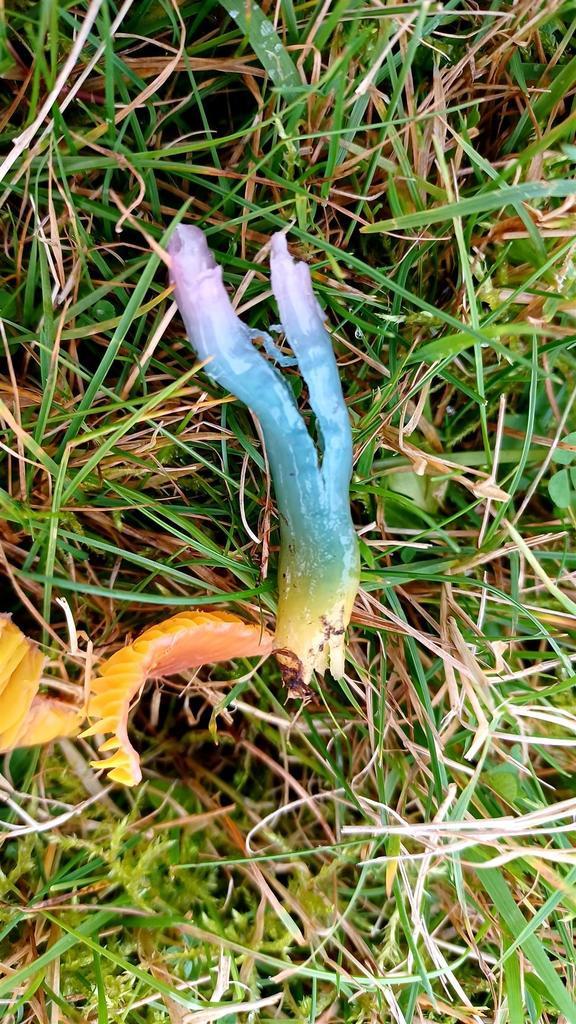
A rainbow of waxcaps
RAINBOW MUSHROOMS!! AAAAAAAAAAAAAAAAAAAA
IT IS SO COOL!!!


Most of these are Parrot Waxcaps or Gliophorus psittacinus, I want to say all of them are but it is a bit difficult to find multiple references that back up a colour range this wide of Parrot Waxcaps. I did find references that back up that Gliophorus psittacinus can be red, orange, yellow and green of colour, but I am not so sure about the blue and purple hues.
Images
1.

2.

3.

4.

5.

6.

7.

8.
9.
~~~~~~~~~~~~~~~~~~~~~~~~~~~~~~~~~~~~~~~~~
Mutuals
@squidsandthings
@fungus-gnats
@fairy-tales-of-yesterday
@flamingears
@lameotello
@lovelyalicorn
@writingraccoon
@edukincon
@emmakapla
Parrot Waxcap - Gliophorus psittacinus
I made this post in reaction to this poll.


Fruitbody
The cap is 5 to 40 milimeters across and is umbonate in shape, expanding to broadly convex or nearly flat. The cap is bald and slimy. It is variable in colours but is most frequently a dark green at first, after which it fades to a orangish yellow from the center outward, till it finallly turns to a dull orangish yellow. The margin of the cap is often thinly lined.¹
The gills are narrowly attached to the stipe; close or nearly distant; adnate. Initially, they usually have a pale green colour, becoming yellowish to orange-yellow throughout development.¹
The stipe is 10 to 80 milimeters long and 2 to 5 milimeters across.² Its surface is bald and slimy. Its pale green above and orangish yellow near its base when young, fading to pale yellowish overall.¹
Spores and microscopic features
The spore print is white in colour.¹
Microscopically, the spores are 6-9 x 3.5-4.5 µ in size, smooth, ellipsoid, hyaline and multiguttulate in KOH, and inamyloid. The basidia are 35-45 µ long, 4-sterigmate or occasionally 2-sterigmate.¹

Ecology and distribution
The precise ecological role uncertain, however, they appear in hardwood, conifer forests and grasslands growing scattered to gregariously. They are frequently found in moss, or on mossy embankments along wooded roadsides. They generally can be found from spring through Autumn.¹
Gliophorus psittacinus can be found in western Europe, the United Kingdom, Iceland, Greenland, the Americas, South Africa and Japan.²
~~~~~~~~~~~~~~~~~~~~~~~~~~~~~~~~~~~~~~~~~
References
1.
2.

~~~~~~~~~~~~~~~~~~~~~~~~~~~~~~~~~~~~~~~~~
Images
1.

2.

3.
~~~~~~~~~~~~~~~~~~~~~~~~~~~~~~~~~~~~~~~~~
Mutuals
@squidsandthings
@fungus-gnats
@fairy-tales-of-yesterday
@flamingears
@lameotello
@lovelyalicorn
@writingraccoon
@edukincon
@emmakapla
I am sorry to hear about Champi getting ''bleached'' and getting kicked out of the company soon, it is a scandal! Horrible and egregious! However I am afraid that it might not be a very good idea to bring a saprobic mushroom into your home.
As much as I want Champi to make it, it is a risky move to take them home.
First of all, if Champi surives being taken home and being brought into a new environment they might release spores as a way to reproduce. Which means the fungus can spread through your home if there is any decaying or dead organic material. Which can be a huge problem.
Even if your were to remove the fruit bodies the mycelium could still be growing anywhere in dead or decaying organic material, so you would need to use proper fungicide/mycocide which can be expensive especially if it is a hardy fungus. Of course in this case I am describing the worst case scenario, I do not mean to cause worry.
HOWEVER, if you can take the piece of furniture, in which the mycelium has grown, home with you and can put it in a controlled space (akin to some kind of lab or a mini-lab, maybe in a securely closed plastic bag depending on the size of the piece of furniture) you might be able to grow it and care for it, but that can still be difficult and expensive.
Shortly, there are definitely risks if you take Champi home with you. You can minimise the risks by growing them in a controlled space but that can be expensive, just like that growing them in an open space and risking an infection can be expensive.
It would be cheaper to leave Champi be, but if you have enough motivation and you can afford to care for them, I suppose you could take them home. If you cannot care for them, you could cut of a piece of the fruitbody and if it has gills you could make a spore print to remember Champi by:)
A tip for making a spore print: if you make it on paper, set the spores with hairspray.
I must say that I am not entirely sure how easily this fungus reproduces and spreads because I am still uncertain of which species it is.
If you have any further questions let me know:)
I am curious to know what will happen to Champi.
~~~~~~~~~~~~~~~~~~~~~~~~~~~~~~~~~~~~~~~~~
@belis86 Another part to the mushroom saga and Champi lore:)
In my workplace there's a mushroom? Fungi? Coworker? So im keeping check on its growth, since I have to maintain my Sanity in hell somehow. Their name is 'Champi'

In case someone know what this fellow coworker is, any information is apreciated.
They grow the white parte first, then the brown parte grows and the other gets small, and so on.
History of my mushroom coworker
Some months ago I thought it was a piece of wood on the really broken furniture, but then it kept growing so hold on its not wood, then it got bigger and had three pointy ends, and had spots, but then the cleaning lady wiped it with bleach, and they "died".
But now they are back, and there is a Lot More, so here se ate documenting it lol
Black elfin saddle - Helvella lacunosa
Another post for the spooky season . . . the black elfin saddle's chambered stipe gives it a skeletal and a somewhat spooky appearance. It might be found in burnt or disturbed grounds.


Fruitbody
The cap is 2 to 4 centimetres across and about 4 to 10 centimetres high.¹ The saddle-shaped cap is irregularly lobed and convoluted, attached to the stipe in several places and has a black to dark brown colour.² It is the smooth outer surface of the cap that bears spores, while the inner surface is sterile.¹
The stipe is 1.5 to 4 centimeters long and 5 to 15 millimetres thick. It might have a whitish colour when young, but soon turns a gray to dark gray colour. The stipe is deeply and ornately ribbed and pocketed, the ribs are rounded, or sometimes sharp and double-edged.²
The flesh of the fruitbody is thin, brittle and chambered.²
Microscopic features
The asci (seen on the bottom left) are typically 340 μm long x 16 μm diameter, each ascus contains eight spores. The paraphyses are about 5 μm diameter, cylindrical¹ and hyaline.² Some paraphyses have capitata apices.¹
The spores (seen on the bottom right) are smooth², ellipsoidal, 15 - 19 x 10 - 13 μm and hyaline. The spore print is white.¹


Ecology
Black elfin saddles can typically be found among leaf litter in all types of woodland, they are often found on burnt ground or in otherwise disturbed woodland clearings.¹ The species is probably mycorrhizal, growing alone, scattered or gregariously.² Helvella lacunosa grows between summer and autumn.¹
Distribution
This species can be found throughout Britain and the throughout the mainland of Europe, it can also be found in North America.¹

Note: the links to the images are in the image discription.
References
1.
2.

Mutuals
@squidsandthings
@fungus-gnats
@fairy-tales-of-yesterday
@flamingears
@lameotello
@lovelyalicorn
@writingraccoon
@edukincon
@emmakapla
White elfin saddle - Helvella crispa
The white elfin saddle is visually the polar opposite twin of the black elfin saddle (discussed in this post), it has a ghost- and skeleton-like appearance.


Fruitbody
The cap is typically 3 to 8cm across and 1 to 4cm high. The saddle-shaped cap is irregularly lobed and has a cream to a pale ochre colour. The upper surface is smooth¹, while the underside is pale ochre and very finely fuzzy. The cap does not attach to the stem where they meet.²
The stipe is 2 to 4 centimetres in diameter and 4 to 8 centimetres long.¹ The stipe is robust, densely ribbed, chambered and have a white to pale yellow colour.²
The flesh of the white elfin saddle is very thin and brittle.²
Microscopic features
The asci (seen on the left below) are typically 300 x 18 μm. Each ascus contains eight spores.¹
The spores (seen on the right below) are ellipsoidal, 18-20 x 10-13 µm and hyaline. The spore print is white.¹


Ecology
Helvella crispa is probably mycorrhizal and can be found growing singularly or gregariously under broadleaf trees, particularly under beeches and oaks, and very often beside paths. It most often emerges from the ground but might also grow from humid hardwoods, meaning it.¹
This mushroom grows from the late summer into the autumn. Sometimes it appears in the winter in warmer regions.²
Distribution
This species can be found growing decidious woodlands in Britain and Ireland, the mainland of Europe and many parts of North America.¹

Note: the links to the images are in the image discription.
References
1.
2.


Mutuals
@squidsandthings
@fungus-gnats
@fairy-tales-of-yesterday
@flamingears
@lameotello
@lovelyalicorn
@writingraccoon
@edukincon
@emmakapla
Candy apple bolete - Exsudoporus frostii
I made this post in response to this poll.


Fruitbody
The cap is 3.5 ¹ to 15 ² centimetres across and initially convex shaped, later becoming broadly convex. The bright red, bald cap is sticky when fresh and its margin is often pale yellow and overhanging slightly.¹
The pore surface is dark red, becoming dirty brownish red and sometimes becoming faintly yellowish near stem and cap margin. The underside of the cap has 2 to 3 circular pores per millimetre at maturity, these tubes are up to 1 centimetre deep.¹ In young specimens yellow droplets can be seen leaking from the pores.³
The stipe is 4 ¹ to 12 ² centimetres long and 1 to 1.5 centimetres thick. It is coarsely reticulate overall with a red reticulum, yellow underneath the red. The basal mycelium is whitish.¹
The flesh is whitish to pale yellow overall and red in the stipe base, turning pale blue when sliced.¹
Microscopic features and spore print
The spores (seen below) are 13–22 x 4–5 µm, fusoid and smooth. The hymenial cystidia are about 40 x 10 µm, fusiform or lageniform, smooth, thin-walled and hyaline in KOH.¹
The spore print is olive brown.¹

Ecology
This species is mycorrhizal with oaks and other hardwoods, growing alone, scattered, or gregariously. It can be found in summer and fall.¹
Distribution
It is widely distributed in Eastern North America, Southwest America, Mexico, and Central America.¹

References:
1.
2.

3.

Mutuals
@squidsandthings
@fungus-gnats
@fairy-tales-of-yesterday
@flamingears
@lameotello
@lovelyalicorn
@writingraccoon
@edukincon
@emmakapla
Beefsteak mushroom - Fistulina hepatica
Which slasher got to this mushroom? Michael Myers? Ghostface?


Fruitbody
The cap is 7 to 20 centimetres across and 7 to 14 centimetres deep, it is irregularly shaped but often semicircular, fan-shaped, or tongue-like, with a lobed and wavy margin. The surface is wet and sticky when fresh, finely bumpy and bald. The cap has a liver red, reddish orange, or brownish red colour.¹
The pore surface is white to pale pinkish in colour, becoming yellowish and eventually reddish brown in age, bruising brown. The tubes are up to 1.5 centimetres long and distinctly separated with circular mouths.¹ This is unlike most polypores in which the pores are bonded together.²
The stipe is absent, or rudimentary and lateral. It is colored like the cap above, covered with the pore surface and firm in texture.¹
The flesh is whitish, streaked with reddish areas. It has a thick, soft and watery texture, exuding a reddish juice when squeezed.¹
Microscopic features and spore print.
The spores (seen below) are ovoid, smooth and about 4.5-6 x 3-4µm.² The spores are also hyaline to yellowish in KOH. The basiadia are 4-sterigmate, the hyphal system is monomitic and clamp connections are present.¹
The spore print is pale pinkish to yellow in colour.²

Ecology
This species saprobic and sometimes weakly parasitic on the wood and deadwood of oaks and other hardwoods, causing a brown rot. Fistulina hepatica is annual, growing from summer to fall. It grows alone or in small groups near the bases of trees and on stumps.¹
Distribution
It is widespread throughout Britain and Ireland and is found throughout the mainland of Europe and is widely distributed throughout North America.²

Note: the links to the images are in the image discription.
References
1.
2.
Mutuals
@squidsandthings
@fungus-gnats
@fairy-tales-of-yesterday
@flamingears
@lameotello
@lovelyalicorn
@writingraccoon
@edukincon
@emmakapla
I forgot to mention, this species of fungi is actually edible when young.
Beefsteak mushroom - Fistulina hepatica
Which slasher got to this mushroom? Michael Myers? Ghostface?
Fruitbody
The cap is 7 to 20 centimetres across and 7 to 14 centimetres deep, it is irregularly shaped but often semicircular, fan-shaped, or tongue-like, with a lobed and wavy margin. The surface is wet and sticky when fresh, finely bumpy and bald. The cap has a liver red, reddish orange, or brownish red colour.¹
The pore surface is white to pale pinkish in colour, becoming yellowish and eventually reddish brown in age, bruising brown. The tubes are up to 1.5 centimetres long and distinctly separated with circular mouths.¹ This is unlike most polypores in which the pores are bonded together.²
The stipe is absent, or rudimentary and lateral. It is colored like the cap above, covered with the pore surface and firm in texture.¹
The flesh is whitish, streaked with reddish areas. It has a thick, soft and watery texture, exuding a reddish juice when squeezed.¹
Microscopic features and spore print.
The spores (seen below) are ovoid, smooth and about 4.5-6 x 3-4µm.² The spores are also hyaline to yellowish in KOH. The basiadia are 4-sterigmate, the hyphal system is monomitic and clamp connections are present.¹
The spore print is pale pinkish to yellow in colour.²
Ecology
This species saprobic and sometimes weakly parasitic on the wood and deadwood of oaks and other hardwoods, causing a brown rot. Fistulina hepatica is annual, growing from summer to fall. It grows alone or in small groups near the bases of trees and on stumps.¹
Distribution
It is widespread throughout Britain and Ireland and is found throughout the mainland of Europe and is widely distributed throughout North America.²
Note: the links to the images are in the image discription.
References
1.
2.
Mutuals
@squidsandthings
@fungus-gnats
@fairy-tales-of-yesterday
@flamingears
@lameotello
@lovelyalicorn
@writingraccoon
@edukincon
@emmakapla